Stem Cell Therapy for Bone Fractures
Stem Cell Therapy for Bone Fractures
Stemcell Consultancy offers stem cell treatments that accelerate healing in bone fractures. Mesenchymal stem cells repair broken tissue, reduce pain, and support fast, strong bone healing.
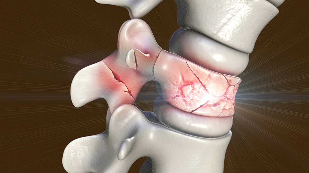
Bone is the body's most prolific self-healer. Break it, and under the right conditions — stable fixation, adequate blood supply, sufficient biological activity at the fracture site — it will rebuild itself over weeks and months, restoring mechanical integrity that no synthetic material can match. This capacity is one of medicine's great conveniences.
The problem is the exceptions.
Between 5 and 10 percent of all fractures fail to heal as expected. Some become delayed unions — fractures that progress too slowly, missing normal healing milestones. Others become nonunions — fractures that stop healing entirely, leaving a biological impasse that requires intervention. For the patients in this category, the implications are significant: prolonged immobilization, persistent pain, functional limitation, multiple surgeries, and in some cases, permanent disability.
Stem cell therapy has emerged as one of the most promising biological tools available to address this failure — not by replacing the surgical management of difficult fractures, but by restoring the osteogenic capacity that makes fracture healing possible in the first place.
Why Some Fractures Don't Heal
To appreciate what stem cell therapy offers, it helps to understand what distinguishes a fracture that heals from one that doesn't.
Normal fracture healing is a sequence of well-orchestrated biological events. After injury, a hematoma forms at the fracture site — the first scaffold. Inflammatory cells arrive, followed by the migration of mesenchymal stem cells from the periosteum, bone marrow, and surrounding soft tissue. These cells differentiate into chondroblasts and osteoblasts, laying down cartilage and then bone across the fracture gap. Callus formation progresses, mineralization occurs, and over months the fracture consolidates into mature bone.
When this process fails, it is almost always due to one or more of the following:
Biological insufficiency — The local pool of osteoprogenitor cells is depleted or dysfunctional. This is more common in elderly patients, in smokers, in patients with diabetes, osteoporosis, or chronic corticosteroid use, and in atrophic nonunions where the biological environment has become quiescent.
Mechanical instability — Excessive movement at the fracture site disrupts the fragile cellular architecture of early callus formation. Even biologically capable bone cannot consolidate in an unstable mechanical environment.
Vascular compromise — Fractures in areas of poor vascularity — the femoral head, scaphoid, talus, certain diaphyseal locations — receive inadequate blood supply to sustain the cellular activity required for healing.
Large bone defects — Fractures with significant bone loss (from high-energy trauma, infection, or tumor resection) exceed the critical defect size beyond which spontaneous regeneration is impossible.
Failed prior intervention — A fracture that has already undergone one or more surgical attempts may have compromised vascularity, scar tissue inhibiting progenitor cell migration, and a biologically exhausted environment.
Stem cell therapy addresses the biological dimension of this problem — specifically, the deficit in osteoprogenitor cells and the impaired anabolic signaling that prevents healing from proceeding.
The Cellular Mechanism of Bone Repair
Mesenchymal stem cells (MSCs) are the primary mediators of bone regeneration. In normal fracture healing, they migrate from periosteum and bone marrow to the fracture site, where they differentiate into osteoblasts — the cells that synthesize bone matrix — and chondroblasts, which form the cartilage template of the fracture callus.
When introduced therapeutically, MSCs work through three overlapping mechanisms:
Direct osteogenic differentiation
Transplanted MSCs integrate into the fracture environment and differentiate into osteoblasts, contributing directly to new bone matrix synthesis. In atrophic nonunions — where the resident progenitor cell population has become exhausted — this direct cellular contribution is the primary therapeutic mechanism.
Paracrine osteoinductive signaling
MSCs release a broad spectrum of growth factors — bone morphogenetic proteins (BMP-2, BMP-7), transforming growth factor-beta (TGF-β), fibroblast growth factor (FGF), vascular endothelial growth factor (VEGF), and platelet-derived growth factor (PDGF) — that activate the fracture site's own resident progenitor cells, stimulate osteoblast proliferation, promote angiogenesis (new blood vessel formation critical for callus vascularization), and drive bone matrix deposition. This paracrine amplification means that introduced MSCs produce a healing response far larger than their numbers alone would suggest.
Anti-inflammatory modulation
Chronic low-grade inflammation is a feature of established nonunions — a biological environment that suppresses osteogenesis and perpetuates tissue quiescence. MSCs modulate this environment by downregulating pro-inflammatory cytokines and shifting the local immune environment toward one that supports regeneration rather than inhibiting it.
The net result: a fracture site that had biologically stalled is provided with the cellular machinery to resume healing.
The Clinical Evidence
The evidence for MSC therapy in fracture healing is the most mature in the orthopedic regenerative medicine field — with decades of preclinical data, multiple clinical trials, and now a comprehensive meta-analysis providing population-level outcome data.
Cui et al. Meta-Analysis — 21 Studies, 866 Patients (BMC Musculoskeletal Disorders, March 2025)
The most comprehensive and current synthesis of the clinical evidence, this systematic review and meta-analysis searched PubMed and Scopus through October 2024, ultimately including 21 studies with 866 patients receiving MSC therapy for nonunion fractures.
The healing rate data is the most clinically meaningful output:
- 44% bone healing at 3 months
- 73% bone healing at 6 months
- 90% bone healing at 9 months
- 91% bone healing beyond 12 months
MSC therapy — whether delivered alone or combined with scaffold materials — was consistently associated with higher odds of bone union and reduced time to union compared to standard treatment. The benefit was particularly pronounced in older patients, where diminished endogenous osteoprogenitor activity makes biological augmentation most valuable. No significant safety concerns were identified across the pooled dataset.
Gomez-Barrena et al. — BM-MSCs with Synthetic Bone Substitute in Long Bone Nonunion
An important clinical study by Gomez-Barrena and colleagues combined bone marrow-derived MSCs with a synthetic bone substitute (MBCP+, comprising 20% hydroxyapatite and 80% beta-tricalcium phosphate) and injected the composite into 28 patients with long bone delayed union or nonunion fractures. At one-year follow-up, 25 of the 28 patients showed bone consolidation on radiographic assessment — a 89% healing rate in a patient population that had already failed standard management, demonstrating the capacity of MSC therapy to rescue biologically stalled fractures.
REBORNE Project — European Multicenter Experience
The REBORNE (Regenerating Bone Defects Using New Biomedical Engineering Approaches) project was a large European multicenter effort that evaluated MSC-based cell therapy combined with biomaterial scaffolds in patients with long bone nonunions and critical-size bone defects. The project produced clinical data across multiple fracture types and locations, confirming both the safety and the biological plausibility of MSC-augmented bone repair in a regulated, multicenter clinical trial framework.
Prophylactic MSC Injection — Randomized Controlled Trial in Distal Tibial Fractures
A Phase I randomized controlled trial evaluated the safety of early prophylactic MSC injection — percutaneous delivery of bone marrow-enriched MSCs into surgically treated distal tibial fractures at 3–6 weeks post-injury — before delayed union had developed. The trial enrolled 24 patients (12 intervention, 12 control) and reached its primary safety endpoint without complications. The biological rationale — that early MSC augmentation could prevent delayed or nonunion rather than treating it after it develops — represents a forward-thinking application with significant potential clinical implications.
Cell Sources and Delivery Methods
Bone Marrow Aspirate Concentrate (BMAC)
The most widely used approach in clinical practice. Bone marrow is aspirated from the posterior iliac crest under local anesthesia, concentrated via centrifugation to enrich the MSC and osteoprogenitor fraction, and injected at the fracture site — often as a same-session procedure. BMAC is autologous (using the patient's own cells), eliminating immune rejection, and can be performed without specialized laboratory infrastructure.
Cultured BM-MSCs
Bone marrow-derived MSCs are harvested, culture-expanded over 2–4 weeks in a cell manufacturing facility, and delivered in substantially higher cell numbers than BMAC provides. The expanded cell product offers greater osteogenic potency but requires a waiting period and more complex logistics. This approach is used in most clinical trial protocols and is more common in academic and specialized centers.
Adipose-Derived Stem Cells (ADSCs)
MSCs harvested from adipose tissue via lipoaspiration. ADSCs are more abundant than bone marrow-derived MSCs per unit volume of tissue harvested, and the harvest procedure is less invasive. Clinical evidence for ADSCs in fracture healing is less extensive than for BM-MSCs but demonstrates comparable biological mechanisms and is accumulating.
MSC-Scaffold Composites
For critical-size bone defects — where a structural void exists that cells alone cannot bridge — MSCs are combined with osteoconductive scaffold materials (hydroxyapatite, beta-TCP, demineralized bone matrix, or bioactive ceramics) that provide a three-dimensional template for bone ingrowth. The scaffold supports cell attachment and proliferation; the MSCs provide the osteogenic and angiogenic drive. This combination approach is most commonly used for large defect reconstruction.
Delivery routes depend on fracture type and clinical context: percutaneous injection under fluoroscopic guidance is the least invasive option for closed fractures; open surgical delivery allows precise scaffold placement in complex reconstructions; and intraoperative delivery at the time of index fixation is used in prophylactic protocols.
Who Should Consider This Treatment?
Stem cell therapy for bone fractures is most clinically appropriate in the following settings:
Established nonunion — Fractures that have failed to progress radiographically for at least three months despite adequate fixation and biological conditions. Atrophic nonunion, where the biological environment is quiescent, is the most compelling indication.
Delayed union — Fractures showing insufficient healing progression at expected milestones, particularly in patients with risk factors for healing impairment (age, diabetes, smoking, osteoporosis, corticosteroid use).
High-risk fractures — Specific fracture locations and patterns known for poor biological healing: scaphoid nonunion, femoral head fractures, tibial diaphyseal fractures in high-risk patients, and fractures with significant soft tissue compromise.
Critical-size bone defects — Post-traumatic bone loss, infection-related debridement defects, or tumor resection gaps that exceed the body's capacity for spontaneous regeneration and require a biological scaffold-cell construct to bridge.
Revision surgery augmentation — In patients undergoing repeat surgical fixation for failed fractures, concurrent MSC delivery enhances the osteogenic environment and improves the biological prospects of the revision procedure.
The Treatment Process
Step 1 — Comprehensive Fracture Assessment Plain radiographs, CT scan for detailed structural assessment of the fracture gap and callus formation, and MRI where soft tissue or vascular assessment is needed. Classification of nonunion type (atrophic, hypertrophic, oligotrophic) guides the biological strategy. Patient-level risk factors — comorbidities, smoking status, medication review — are identified and optimized where possible before biological intervention.
Step 2 — Mechanical Stability Confirmation Stem cell therapy addresses the biological dimension of nonunion. It will not overcome mechanical instability. If fixation is inadequate — implant failure, loss of reduction, excessive fracture site motion — mechanical revision must precede or accompany biological augmentation. A nonunion with adequate fixation and biological insufficiency is the ideal indication; a nonunion with both mechanical and biological failure requires both to be addressed.
Step 3 — Cell Harvest and Preparation For BMAC: same-day aspiration from the iliac crest, centrifugation, and concentration — completed in 30–45 minutes under local anesthesia. For cultured MSCs: bone marrow or adipose harvest followed by 2–4 weeks of laboratory expansion before delivery. Cell count, viability, and sterility testing are performed before clinical use.
Step 4 — Guided Delivery Percutaneous injection under fluoroscopic guidance directly into the fracture gap or nonunion site. For scaffold composites, an open or mini-open approach delivers the MSC-scaffold construct with precise positioning. The injection procedure itself is brief — typically under 30 minutes.
Step 5 — Monitoring and Follow-Up Serial radiographs at 6, 12, and 24 weeks assess callus formation and fracture bridging. CT scan at 3–6 months provides definitive assessment of bone consolidation. Functional rehabilitation is calibrated to healing progress — weight-bearing progression guided by radiographic evidence of union.
Frequently Asked Questions
How is this different from a standard bone graft? Autologous iliac crest bone graft provides osteoconductive scaffold material and some osteoprogenitor cells — and remains a well-established option. MSC therapy, particularly with culture-expanded cells, delivers a substantially higher number of biologically active osteoprogenitor cells and growth factors than a standard bone graft. The 2025 meta-analysis showed MSC therapy associated with higher union rates and shorter union time compared to standard bone graft — though the two can also be combined.
Can MSC therapy avoid the need for repeat surgery? In selected patients with nonunion fractures that have adequate mechanical fixation, percutaneous MSC injection may provide the biological stimulus needed for consolidation without requiring open revision surgery. This is one of the most compelling aspects of the approach — delivering a meaningful biological intervention through a needle rather than an operating theatre.
How long after a fracture can MSC therapy still be effective? The biological environment of a long-standing atrophic nonunion is quiescent but not permanently closed. Clinical studies have documented successful bone consolidation after MSC injection in fractures with nonunion periods ranging from months to years, provided mechanical stability is adequate. Earlier intervention generally produces faster responses.
Is the iliac crest harvest painful? The aspiration procedure is performed under local anesthesia. Most patients describe moderate pressure rather than significant pain during the procedure, with some soreness at the harvest site for a few days afterward. The discomfort is consistently described as well within the tolerable range for an outpatient procedure.
What are the success rates? The 2025 Cui et al. meta-analysis — pooling 21 studies and 866 patients — documented an eventual bone union rate of 91% in patients treated with MSC therapy for established nonunions. Individual outcomes depend on nonunion type, fracture location, mechanical environment, and patient factors — but these population-level numbers represent a clinically meaningful success rate in a patient group that has already failed standard management.
Can this be done at the same time as revision surgery? Yes — intraoperative MSC delivery at the time of revision fixation is a recognized approach that combines mechanical restoration with biological augmentation in a single procedure, avoiding the need for a second intervention.
The Bottom Line
Most fractures heal. The ones that don't — the delayed unions, the atrophic nonunions, the critical defects — represent a disproportionate burden of suffering and surgical intervention. For patients caught in this cycle of incomplete healing and repeated procedures, the availability of a biologically active treatment that can be delivered percutaneously, without major additional surgery, and that achieves bone union in over 90% of cases in pooled clinical data, is a genuinely significant development.
Stem cell therapy for fracture nonunion is not experimental in concept — it is the application of well-understood osteogenic biology to a problem that conventional medicine has long struggled to solve reliably. The evidence, culminating in the 2025 meta-analysis of 866 patients, supports its use as a meaningful addition to the orthopedic toolkit for managing difficult fractures.
Schedule a consultation to discuss whether biological augmentation is appropriate for your fracture situation and what a tailored treatment plan looks like.
This article is for informational purposes only and does not constitute medical advice. Fracture management should always be directed by a qualified orthopedic surgeon.
Quick Question